The IU Light Microscopy Imaging Center (LMIC) was awarded a high-end instrumentation grant to procure an Applied Precision OMX Super Resolution imaging system. Scheduled for installation in mid-July, the LMIC adds this system to its impressive array of microscopes. With its new addition, the LMIC becomes the go-to facility for researchers requiring light microscopy, image processing, and image analysis.
For roughly two years, the LMIC has offered access to multiple imaging systems to researchers on the IUB campus as well as to outside users within the state of Indiana. The facility, located in Myers Hall on the Bloomington campus, is utilized by faculty and graduate students from many disciplines including biology, chemistry, optometry, neurobiology, and medical sciences.
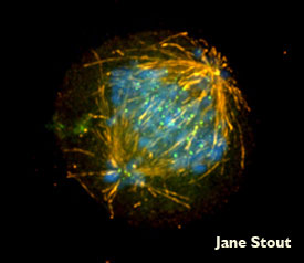
In May, the LMIC was awarded approximately $1.2 million in funding through the American Recovery and Reinvestment Act to purchase an Applied Precision OMX Super Resolution imaging system. Through the use of 3-D structured illumination, this new system allows researchers to resolve sub-cellular structures that were in the past too small to clearly discern. The technology offered with this system will benefit a number of researchers, particularly those studying microbiology, chromatin structure, or any aspects of cell structure.
“Previous microscopes would tell [scientists] whether a protein was in a small bacterial cell,” said Claire Walczak, executive director of the LMIC and professor of biochemistry and molecular biology in Bloomington’s Medical Sciences Program. “The new technology will allow them to know where within a cell different proteins are localized. For chromatin researchers, there is the potential to locate where specific genes are located or expressed within a nucleus. Cell biologists can ask questions about protein localization at unprecedented resolution.”
The new imaging system is expected to produce results that could lead to groundbreaking discoveries and deepen the understanding of cell processes. “The microscope also has the potential to capture 3-D images at near-live speeds to allow researchers to study highly dynamic processes in cells,” Walczak said.
The LMIC also recently unveiled a 64-bit image-processing workstation in early 2010. Technological advances in imaging systems have resulted in vast amounts of data being collected at a greater speed, yet this increase in data has made it more difficult for researchers to analyze and interpret such data. Thus the new workstation, with its implementation of automated algorithms, is an essential tool for researchers to more effectively mine data. The new image processing workstation also provides the capability to rotate and reconstruct images as well as distinguish subcellular structures from varying perspectives. Featured software includes AutoQuant for image deconvolution and Imaris for 3-D image reconstruction, modeling, and presentation.
The LMIC is accessible 24 hours a day to researchers and graduate students that have been properly trained and approved by LMIC staff members Sid Shaw, technical director and assistant professor of biology, and Jim Powers, manager and assistant research scientist.

 The College of Arts